Our Office
28562 Oso Parkway, Suite K
Rancho Santa Margarita,
CA 92688
CA 92688
Existing Patients: (949) 505-9901
New Patients: (949) 272-0877
Fax: (949) 505-9902
Visit Us Online

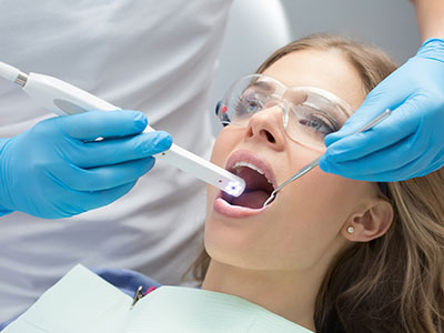
An intraoral camera is a compact, pen-sized imaging tool designed specifically for use inside the mouth. Equipped with a tiny, high-resolution lens and built to capture full-color images in tight spaces, the device gives clinicians a magnified view of teeth, gums, and other oral tissues. Images are displayed in real time on a chairside monitor so both the patient and dental team can view the oral condition together, helping translate what can otherwise be hard-to-see into clear visual information.
Unlike traditional exterior photography, an intraoral camera is optimized for intraoral lighting, angles, and access. Its form factor allows clinicians to inspect occlusal surfaces, interproximal areas, restorations, and soft tissue details without invasive probes or awkward positioning. Because the camera captures crisp images at multiple magnifications, it becomes an effective extension of the clinical exam — offering visual evidence that complements tactile assessment and radiographic findings.
For patients, the immediate visual feedback an intraoral camera provides helps make routine exams more transparent. Seeing an image of a suspected problem or a restorative margin on screen establishes a shared frame of reference for discussing treatment options, expected outcomes, and preventive steps. That combination of clarity and immediacy has made intraoral imaging a standard element of contemporary dental care.
High-resolution intraoral images allow clinicians to identify subtle patterns that might be difficult to detect with the naked eye alone. Small hairline cracks, early enamel breakdown, marginal gaps around existing restorations, and localized soft-tissue irritation are often more evident when magnified and captured for review. These visual cues, when combined with a thorough clinical exam and radiographs, strengthen diagnostic accuracy and support more confident treatment planning.
Because intraoral cameras produce reproducible, detailed images, they are particularly useful for monitoring change over time. Dentists can compare current images with earlier captures to track lesion progression, restoration wear, or periodontal tissue response to therapy. This chronological record helps clinicians decide whether a conservative approach, additional diagnostics, or restorative intervention is appropriate, and it reduces uncertainty in borderline cases.
Intraoral photography can also reduce the risk of missed findings during complex procedures. When treating multi-surface cavities, planning restorative margins, or assessing margins of crowns and veneers, real-time visualization helps the clinician verify clinical endpoints before finalizing treatment. That extra layer of precision contributes to predictable outcomes and minimizes the need for unnecessary adjustments later.
One of the most practical benefits of intraoral cameras is their role in patient communication. Rather than relying solely on verbal descriptions, clinicians can show patients the actual problem: a dark spot between teeth, inflammation at the gumline, or a small fracture that could grow larger. This visual evidence makes explanations easier to follow and helps patients understand why a recommended procedure may be in their best interest.
When patients see clear images of their own mouths, they typically ask more informed questions and participate more actively in treatment discussions. The camera supports a collaborative approach, enabling the dental team to walk patients through anatomy, demonstrate how proposed treatments will address specific concerns, and outline preventive measures to maintain oral health. This shared understanding often leads to better adherence to home care and follow-up appointments.
Patient-focused imaging also supports informed consent. Presenting clinical findings visually allows patients to review the exact areas of concern and grasp potential risks and alternatives. That transparency strengthens trust between the patient and provider while ensuring treatment decisions are based on clear, mutually understood information.
Intraoral cameras integrate smoothly into modern digital dental records, enabling clinicians to store, organize, and retrieve images as part of each patient’s chart. These images serve as an objective clinical record that can be referenced during future visits, treatment planning sessions, or discussions with other members of the dental team. Well-documented imagery improves continuity of care, particularly in cases that require follow-up over months or years.
When collaboration with specialists or dental laboratories is necessary, high-quality intraoral photos can be shared to clarify clinical findings and streamline communication. Clear visuals help specialists understand the referring clinician’s concerns, while labs benefit from detailed images when fabricating restorations that must match contours, shade relationships, or occlusal anatomy. This reduces ambiguity and can speed the planning and fabrication process without compromising clinical standards.
Storing images digitally also supports compliance and quality management. Having a visual baseline for restorative work, periodontal status, or mucosal conditions makes it easier to audit outcomes, document informed consent, and support clinical notes. The convenience of retrieving images during chairside consultations makes them a practical tool for both everyday exams and complex case management.
Using an intraoral camera is minimally invasive and generally well tolerated by patients of all ages. The slim handpiece slips comfortably into the oral cavity, and captures are quick — most images are taken in seconds. Because the process avoids repeated probing or prolonged discomfort, it can be particularly helpful for anxious patients, children, or individuals with gag reflex sensitivity who benefit from a less intrusive diagnostic approach.
Clinically, best practices include using disposable sheaths or barriers to maintain strict infection control, positioning the camera for optimal illumination and focus, and combining image capture with a targeted clinical exam. Technicians and hygienists trained in intraoral photography can acquire consistent, reproducible images that make interpretation easier for the dentist and more meaningful for the patient.
At Rise & Shine Dental Group, intraoral imaging is part of a broader commitment to accurate diagnosis, clear communication, and patient comfort. We use digital tools in ways that complement clinical expertise, safeguard patient safety, and support efficient treatment planning while maintaining a focus on personalized care.
In summary, intraoral cameras bring real-time, magnified images into the dental exam — improving diagnosis, enhancing patient communication, and supporting a modern digital workflow. If you’d like to learn more about how we use intraoral imaging in routine exams or specific treatments, please contact us for more information.
An intraoral camera is a small, pen-sized imaging device designed to capture high-resolution, full-color photos of teeth and soft tissues inside the mouth. Its miniature lens and integrated illumination allow clinicians to photograph occlusal surfaces, interproximal areas and restoration margins that are difficult to see with the naked eye. Images appear in real time on a chairside monitor to supplement tactile exams and radiographic findings.
Many systems offer multiple magnifications and capture modes to document subtle cracks, early enamel breakdown and soft-tissue findings. Captures are stored digitally in the patient record so clinicians can review and compare them during treatment planning. Because the camera is noninvasive and quick to use, it fits naturally into routine checkups and restorative visits.
High-resolution intraoral images reveal surface details that are often missed by unaided vision, including hairline fractures, marginal gaps around restorations and focal enamel defects. These visual cues strengthen the clinical examination by providing objective, reproducible evidence that complements probing and radiographs. When findings are documented photographically, clinicians can make more confident decisions about whether to monitor, treat conservatively or proceed with restoration.
Because images are reproducible, they are especially valuable for monitoring change over time and reducing diagnostic uncertainty in borderline cases. Comparing current photos with earlier captures helps clinicians detect progression of lesions, restoration wear or soft-tissue response to therapy. This chronological record supports informed treatment planning and can reduce the risk of missed findings during complex procedures.
An intraoral camera exam is generally quick, comfortable and minimally invasive; the slim handpiece slips into the mouth and most photos are captured in seconds. Patients typically view images on a chairside monitor while the clinician explains findings, which helps translate clinical observations into clear visual information. The process avoids prolonged probing and can be easier for patients who have a sensitive gag reflex or dental anxiety.
Clinicians and trained team members position the camera for optimal illumination and focus, and captures are repeated only as necessary to document relevant areas. Disposable sheaths or barriers are used to maintain infection-control standards and the camera is disinfected between patients. Seeing their own dental images usually encourages patients to ask questions and participate more actively in treatment discussions.
Intraoral photographs provide clear, personalized visuals that help patients understand specific conditions in their mouths, such as interproximal decay, gum inflammation or a flawed restoration margin. Visual evidence makes explanations easier to follow than verbal descriptions alone and establishes a shared frame of reference for discussing options. That transparency helps patients weigh risks and benefits and increases clarity during consent conversations.
When patients can see the exact area of concern, they tend to ask more informed questions and feel more confident about recommended next steps. The images also allow the dental team to demonstrate anatomy, explain how proposed treatments will address the problem and outline preventive measures to reduce recurrence. This shared decision-making often leads to better adherence to home care and follow-up recommendations.
An intraoral camera is complementary to radiographs and clinical probing rather than a replacement for them. Cameras excel at documenting surface detail and soft-tissue appearance, while X-rays, CBCT and other imaging modalities reveal internal structure, bone levels and sub-surface pathology. Together, these tools provide a more complete diagnostic picture than any single modality alone.
Clinicians combine visual intraoral images with radiographic findings and clinical tests to confirm diagnoses and plan treatment. For example, a camera can show a visible crack or marginal gap while an X-ray can assess the underlying tooth structure and root. Using both methods improves diagnostic confidence and helps determine the appropriate intervention.
Intraoral images are integrated into electronic dental records and stored as part of each patient’s chart, creating an objective visual record of clinical findings. Modern practice management systems protect these records with standard security measures and access controls to help preserve patient privacy. Clinicians retrieve images during follow-up visits to compare changes and document outcomes over time.
When collaboration is needed, high-quality intraoral photos can be shared with specialists or dental laboratories to clarify clinical concerns and streamline treatment planning. Images are shared only as part of coordinated care and in accordance with the practice’s privacy policies and applicable regulations. Clear visuals improve communication with collaborators and reduce ambiguity in restorative fabrication and specialist consultations.
All patients can benefit from intraoral imaging, but certain groups see particular advantages: patients with complex restorative needs, those undergoing long-term monitoring, individuals who have difficulty visualizing dental issues and people who are anxious about dental care. Children and patients with heightened gag reflexes often tolerate intraoral photography better than repeated probing. Visual documentation also helps patients track progress after periodontal therapy or restorative treatment.
Clinicians and support staff benefit as well, because consistent images improve charting accuracy and streamline communication within the care team. Dental laboratories gain a clearer understanding of occlusal anatomy and shade relationships when photos are provided. Overall, intraoral imaging strengthens diagnosis, patient engagement and multidisciplinary coordination.
Standard infection-control measures include using single-use disposable sheaths or barriers over the camera tip and disinfecting the handpiece between patients according to manufacturer and regulatory guidance. These practices prevent cross-contamination while preserving image quality. Team members are trained to handle the device safely and to follow the practice’s established sterilization and surface-disinfection protocols.
Proper positioning and minimal intraoral contact reduce the need for repeated maneuvers and help maintain a hygienic workflow. Clinicians also combine camera use with routine glove changes and hand hygiene to further reduce risk. Following consistent protocols protects patient safety and supports reliable, reproducible imaging results.
Intraoral cameras are a key component of a modern digital workflow, feeding high-quality images directly into electronic health records, charting modules and case documentation. These images can be paired with digital impressions, radiographs and CAD/CAM data to create comprehensive records that support efficient treatment planning. The ability to attach photos to specific chart entries improves clarity in clinical notes and assists long-term case management.
When working with dental laboratories or specialists, clinicians can include intraoral photos with prescriptions and referral documentation to clarify clinical intent. This reduces back-and-forth communication and helps labs fabricate restorations that better match contours and occlusal anatomy. The result is a more coordinated process from diagnosis through final restoration.
At Rise & Shine Dental Group, intraoral imaging is part of our commitment to accurate diagnosis, clear communication and patient comfort. The technology helps the team identify subtle clinical signs earlier, explain findings visually during chairside consultations and integrate images into the patient’s digital record for ongoing care. Using intraoral cameras supports our focus on evidence-informed treatment planning and predictable outcomes.
Beyond diagnostics, intraoral photography enhances patient education and shared decision-making, which aligns with our mission to provide personalized dental care. The noninvasive nature of the device makes exams more comfortable while improving documentation for follow-up and collaboration. Together with other digital tools, intraoral imaging helps deliver precise, patient-centered dentistry.