Our Office
28562 Oso Parkway, Suite K
Rancho Santa Margarita,
CA 92688
CA 92688
Existing Patients: (949) 505-9901
New Patients: (949) 272-0877
Fax: (949) 505-9902
Visit Us Online

Digital radiography replaces traditional film with electronic sensors and computer processing to capture dental x‑ray images. Instead of waiting for film to be developed, the sensor converts x‑ray exposure into a digital file that appears on a computer screen almost instantly. This shift from chemical processing to digital capture has changed how dentists diagnose conditions, plan treatment, and communicate findings with patients.
For patients, the most noticeable difference is speed and clarity: images are available immediately and can be enhanced for better viewing. For clinicians, digital radiography streamlines recordkeeping and supports more precise diagnosis thanks to tools like contrast adjustment, magnification, and measurement features built into imaging software. Because the images are digital files, they fit seamlessly into electronic health records and can be archived without physical storage concerns.
Adopting digital radiography also supports safer, more efficient care. It eliminates film-processing chemicals and reduces the need for repeat exposures by enabling real-time verification of image quality, which benefits patients and the office environment alike. By modernizing the imaging workflow, dental teams can focus more time on patient care and less on manual processing tasks.
Digital sensors come in several forms, including solid-state intraoral sensors and phosphor plate systems. These technologies capture high-resolution images of teeth, roots, and surrounding bone structures, revealing details that are crucial for diagnosing cavities, periodontal disease, fractures, and other oral conditions. The clarity and consistency of digital images help clinicians detect issues earlier and with greater confidence.
Beyond image resolution, software-driven tools increase diagnostic value. Clinicians can zoom in, adjust brightness and contrast, and measure anatomical features directly on screen. These capabilities make it easier to compare sequential images over time, monitor healing, and evaluate the effectiveness of treatment. The result is a more precise, evidence-based approach to clinical decision-making.
From a patient comfort perspective, many modern sensors are designed with ergonomics in mind. Slimmer sensor profiles and flexible positioning reduce gag reflex triggers and make intraoral imaging more tolerable for a wider range of patients, including children and those with strong gag reflexes. Faster capture times also cut down on how long a patient needs to hold a sensor in place, improving the overall experience.
One of the most important advantages of digital radiography is improved control over radiation exposure. Digital sensors are more sensitive to x‑rays than film, so effective images can often be acquired with less radiation. Additionally, modern systems include exposure settings and automatic exposure control features that help clinicians tailor dose to the diagnostic need and the size of the patient.
Digital imaging also reduces the likelihood of retakes. When an image is captured, the clinician can review it immediately and determine whether it meets diagnostic requirements, correcting positioning or exposure in real time. Fewer retakes mean fewer x‑ray exposures overall, which aligns with the principle of minimizing radiation while still obtaining necessary diagnostic information.
Practices that use digital radiography combine sensor technology with established safety protocols—such as using lead aprons when appropriate, following recommended imaging guidelines, and maintaining equipment—to ensure that patients receive safe, effective care. These layers of protection support routine screenings and targeted imaging with patient safety as a priority.
Digital radiography transforms the way images are stored, retrieved, and shared. Because images are digital files, they can be integrated into patient records and accessed instantly during appointments. This immediacy helps clinicians explain conditions to patients in real time, using the actual images to illustrate findings and discuss treatment options.
Sharing images with specialists or outside providers is also straightforward with digital systems. Secure electronic transfer of radiographs facilitates coordination when referrals or collaborative care are needed, eliminating the delays and hassles of physical film. For complex cases, having high-quality digital images readily available supports better interdisciplinary planning and continuity of care.
For the dental team, digital radiography improves workflow efficiency. Appointments can run more smoothly because imaging steps are faster and administrative overhead is reduced. Staff can focus on patient education and treatment planning rather than on manual processing, which enhances the overall patient experience and helps the practice operate more effectively.
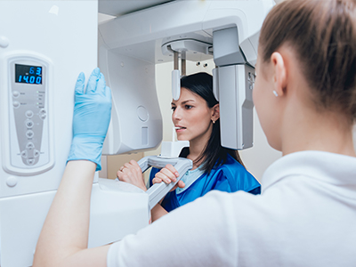
Undergoing digital dental x‑rays is a quick and straightforward process. When intraoral images are needed, a small sensor or plate is positioned in the mouth while the clinician or assistant aligns the x‑ray tube. The exposure itself takes only a fraction of a second, and the resulting image appears on the monitor almost immediately for review. Extraoral digital imaging, used for broader views such as panoramic images, takes only slightly longer and is noninvasive.
Before any x‑ray is taken, the dental team will explain the reason for the imaging and what they hope to learn from the images. Patients are encouraged to mention any concerns—such as pregnancy—so the team can take appropriate precautions. Protective measures like lead aprons may be used according to standard safety practices to further minimize exposure.
After images are captured, the clinician will review them with the patient, pointing out areas of interest and explaining how the findings relate to oral health and treatment planning. Because digital images can be enhanced and enlarged on screen, patients often find it easier to understand their condition and the rationale behind recommended care. If additional imaging or a specialist consultation is needed, the digital files make it simple for the practice to coordinate next steps efficiently.
Digital radiography is a modern, patient-centered approach to dental imaging that delivers faster results, improved diagnostic clarity, and enhanced safety compared with traditional film techniques. By combining high-quality sensors with image-enhancement tools and electronic record systems, digital x‑rays support better clinical decisions and more effective patient communication.
At Rise & Shine Dental Group, our team uses digital radiography as part of a comprehensive approach to diagnosis and treatment planning, always with an emphasis on patient comfort and safety. If you have questions about how digital imaging is used during your visit or what to expect at your next appointment, please contact us for more information.
Digital radiography uses electronic sensors and computer processing to capture dental x-ray images instead of traditional film. The sensor converts x-ray exposure into a digital file that appears on a computer screen almost immediately, which speeds diagnosis and reduces the need for chemical processing. Because images are digital, they can be enhanced, measured, and archived without the physical storage concerns associated with film.
This technology matters because it supports more precise, evidence-based decision making and clearer patient communication. Immediate image availability lets clinicians verify quality in real time and reduce repeat exposures, improving efficiency in the operatory. Integration with electronic health records makes it easier to track changes over time and coordinate care when a specialist consultation is needed.
Digital sensors are solid-state devices or phosphor plates that capture x-rays and produce high-resolution images for on-screen review, while traditional film relies on chemical development to reveal an image. Sensors are more sensitive to x-rays, so they generally require lower exposure to produce diagnostically useful images, and they eliminate the need for developing chemicals and darkroom procedures. The consistency and clarity of digital capture also reduce variability in image quality from one exposure to the next.
Beyond sensitivity, many modern sensors are thinner and ergonomically designed to improve patient comfort during intraoral imaging. Software tools allow clinicians to adjust contrast, magnify areas of interest, and take precise measurements directly on-screen, increasing the diagnostic value of each exposure. These capabilities streamline workflow and allow staff to spend more time on patient care rather than manual processing.
Yes, digital dental x-rays are considered safe when used appropriately; the sensors are more sensitive than film, which often allows for lower radiation doses while still achieving clear diagnostic images. Clinicians follow the ALARA principle—keeping exposure As Low As Reasonably Achievable—by tailoring exposure settings to the diagnostic need and the patient’s size. Routine safety measures such as periodic equipment maintenance, correct positioning, and use of protective shielding when appropriate further reduce risk.
Digital systems also reduce the need for retakes because images are available immediately and can be verified on-screen, which lowers the cumulative radiation a patient receives. Patients who are pregnant or have specific medical concerns should inform the dental team so that additional precautions or alternative imaging approaches can be considered. Overall, modern digital radiography provides a highly controlled method for obtaining necessary diagnostic information with patient safety as a priority.
Digital radiography supports a range of image types, from intraoral bitewings and periapical images that visualize individual teeth and surrounding bone to extraoral panoramic images that show broader jaw relationships. In addition, cone beam computed tomography (CBCT) produces three-dimensional views that are useful for implant planning, complex endodontic assessments, and evaluating anatomy prior to oral surgery. Each modality serves different diagnostic purposes and is selected based on the clinical question at hand.
The choice of imaging type depends on the information needed, the area of interest, and the patient’s history. For routine cavity detection and periodontal assessment, bitewings and periapicals are most common, while panoramic or CBCT images are reserved for comprehensive treatment planning and complex cases. Clinicians combine clinical examination with the appropriate imaging modality to obtain a complete and accurate assessment.
Digital radiographs reveal a wide range of conditions that may not be visible during a clinical exam alone, including interproximal cavities, bone loss from periodontal disease, dental abscesses, impacted or unerupted teeth, root fractures, and changes in the jawbone. High-resolution images help clinicians identify early signs of disease so treatment can be planned before problems advance. Because images can be magnified and enhanced, subtle changes are easier to recognize and document.
Radiographs are also valuable for monitoring treatment outcomes, such as root canal therapy healing or bone integration around implants, by allowing sequential comparison over time. They complement the clinical exam rather than replace it, and findings on an x-ray are interpreted alongside patient history, symptoms, and intraoral evaluation to guide appropriate care. When necessary, additional or specialized imaging can be used to clarify ambiguous findings.
In most cases, no special preparation is required for dental digital x-rays; patients can eat, take medications, and maintain normal oral hygiene before the appointment. You should inform the dental team if you are pregnant or suspect you might be, as the team will take extra precautions and consider postponing nonurgent imaging when appropriate. It is also helpful to mention any recent imaging from other providers so those files can be requested and compared if needed.
On the day of imaging, remove jewelry, eyeglasses, or removable oral appliances that could interfere with image capture. The dental team will explain the procedure, position the sensor or plate comfortably in the mouth, and provide any protective shielding necessary. The exposure itself is very brief, and images are available instantly for review with the clinician.
Clinicians use digital images as a core component of diagnosis and treatment planning by combining visual findings from radiographs with clinical examination and patient history. Software tools enable clinicians to zoom, measure structures, adjust contrast, and compare sequential images, which aids in detecting progression of disease and evaluating the success of therapy. Precise measurements from digital images support restorative planning, endodontic work, and implant placement by clarifying anatomical relationships and dimensions.
Digital imaging also facilitates more conservative and targeted treatment because clinicians can identify the exact location and extent of a problem before intervening. For complex cases, images are used to simulate treatment steps, plan surgical approaches, and consult with specialists while sharing clear visual information. This evidence-based approach improves clinical confidence and helps patients understand the rationale for recommended care.
Digital radiography makes it easier to involve patients in their own care by allowing clinicians to display images on a monitor and explain findings visually during the appointment. Being able to enlarge, highlight, and annotate areas of concern helps patients better understand their condition and the proposed treatment options. This visual approach builds transparency and supports informed decision-making without relying solely on verbal descriptions.
For referrals or collaborative care, digital files can be shared securely with specialists to streamline diagnosis and planning. Electronic transfer reduces delays and preserves image quality, which is important when multiple clinicians need to review the same images. Overall, digital sharing enhances continuity of care and helps ensure everyone involved has access to the same high-quality diagnostic information.
Yes, digital radiographs are stored as electronic files that can be managed within secure practice systems and integrated into electronic health records. Modern dental software supports encrypted backup and controlled access so that images are preserved and only available to authorized users. Secure storage also simplifies recordkeeping and long-term monitoring by eliminating the degradation risks associated with physical film.
When images must be shared with another provider, secure electronic transfer methods are used to protect patient privacy and comply with applicable regulations. Practices maintain protocols for data retention, access control, and secure sharing to ensure that diagnostic images remain confidential while still enabling timely collaboration when specialist input is needed.
At Rise & Shine Dental Group, digital radiography is used as a standard tool to enhance diagnosis, treatment planning, and patient communication while prioritizing comfort and safety. Our team uses high-resolution sensors and image-enhancement software to document oral health, compare images over time, and tailor care to each patient’s needs. Immediate image availability lets clinicians verify quality and discuss findings with patients during the visit, which supports clear, collaborative decision-making.
We combine digital imaging with established safety protocols and individualized assessment to ensure each exposure is justified and optimized for the diagnostic task. Digital files are integrated into the patient record for continuity of care and can be shared securely with specialists when collaborative treatment is necessary. This approach helps us deliver precise, efficient, and patient-centered care at every visit.