Our Office
28562 Oso Parkway, Suite K
Rancho Santa Margarita,
CA 92688
CA 92688
Existing Patients: (949) 505-9901
New Patients: (949) 272-0877
Fax: (949) 505-9902
Visit Us Online

At Rise & Shine Dental Group, we combine clinical experience with advanced imaging to deliver clear, reliable diagnostic information. Cone‑beam computed tomography (CBCT) gives clinicians a three‑dimensional view of dental and facial structures that traditional two‑dimensional X‑rays cannot provide. When used thoughtfully, CBCT helps clinicians diagnose complex conditions more accurately and design treatment plans that are tailored to each patient’s anatomy.
Our Rancho Santa Margarita office uses CBCT as part of a comprehensive imaging strategy — not as a routine substitute for standard X‑rays. The goal is always to gather the specific information needed to guide care while keeping procedures comfortable and efficient for patients. Below are the key ways CBCT enhances diagnosis, planning, and patient communication.
Cone‑beam CT captures volumetric images in a single, brief scan, producing a stack of cross‑sectional slices that can be viewed from any angle. This 3D perspective reveals relationships between teeth, jawbone, nerves, and sinuses that are often hidden on flat images. Clinicians can inspect bone contours, tooth roots, and soft tissue outlines with far greater confidence than is possible with traditional radiographs.
Because the images are volumetric, clinicians can measure distances and angles directly on the scan, which reduces uncertainty when evaluating complex anatomy. This precision is particularly helpful in cases where overlapping structures or atypical anatomy might obscure important details on standard films. In short, CBCT converts uncertainty into actionable information.
Another diagnostic advantage is the ability to detect subtle changes in bone density, pathologic lesions, or anatomic variations that could affect treatment. Identifying these issues early — and understanding their exact location and extent — allows for more predictable, safer treatments and clearer conversations with patients about options and expectations.
CBCT provides a comprehensive view of the maxillofacial region, including the teeth, alveolar bone, temporomandibular joints, nasal cavity, and paranasal sinuses. In endodontics, clinicians can locate unusual root canal anatomy, detect previously missed canals, and visualize periapical pathology in three dimensions. This level of detail can change the diagnosis or indicate a different approach to treatment.
For periodontal assessment, CBCT helps quantify bone loss and fenestrations, and it can clarify the extent of defects around compromised teeth. When evaluating trauma, the scan can reveal root fractures, displacement of teeth, and subtle cortical disruptions that would be difficult to appreciate on planar X‑rays. The ability to visualize these conditions helps clinicians recommend treatments that are anatomically appropriate.
CBCT is also useful for airway analysis and sleep‑related breathing evaluations, where anatomic constrictions are implicated. While a scan alone does not replace a full medical assessment, it gives valuable structural information that supports a multidisciplinary approach to airway and sleep concerns. Overall, the scan’s versatility makes it a powerful diagnostic tool across many areas of dental care.
One of the most transformative uses of CBCT is in implant dentistry. Accurate assessment of bone volume, ridge morphology, and the location of critical structures such as the inferior alveolar nerve or the maxillary sinus is essential to predictable implant placement. CBCT enables clinicians to visualize the proposed implant site in three dimensions and select implant size and angulation with greater certainty.
Many practices use CBCT data to perform virtual treatment planning, which can be carried into the operatory through surgical guides or real‑time navigation. This workflow reduces guesswork during surgery, supports minimally invasive approaches, and improves the likelihood of long‑term implant success. For patients, that often means shorter procedures and more predictable healing.
CBCT also plays a key role in other surgical procedures, such as impacted tooth removal, assessment of cysts or lesions, and evaluation prior to orthognathic surgery. By mapping anatomy precisely before entering the operating field, clinicians can anticipate challenges and coordinate care with specialists when necessary, improving safety and outcomes.
Radiation exposure is an important consideration with any imaging modality. Modern CBCT units are designed to minimize dose while delivering clinically useful images. Protocols can be adjusted for the area of interest and the patient’s size so that only the necessary volume is scanned. When indicated, a limited‑field scan targets the specific region of concern rather than imaging the entire jaw.
Compared with medical CT, many dental CBCT systems use lower radiation levels while still providing fine bony detail suitable for dental applications. Clinicians follow the ALARA principle — keeping exposure "as low as reasonably achievable" — and order scans only when the additional information will influence diagnosis or treatment decisions. This responsible approach balances diagnostic benefit with patient safety.
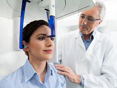
Patient comfort is also a priority. CBCT scans are fast — typically completed in under a minute — and require minimal repositioning. The open design of most dental CBCT units reduces feelings of confinement and allows for easier accommodation of patients who may have mobility limitations. Clear communication about the process helps patients feel comfortable and reassured before imaging begins.
Finally, our team reviews each scan carefully and integrates the findings into a clear treatment plan. If a scan reveals information outside the scope of dental care, we coordinate with medical providers as needed to ensure a comprehensive approach to the patient’s health.
Beyond diagnosis and surgical planning, CBCT enhances how clinicians personalize care. Three‑dimensional images allow for precise measurements and simulations that can be discussed with patients during consultations. Visualizing anatomy in 3D helps patients understand the reasons behind recommended treatments and the steps involved in care.
CBCT data can be combined with intraoral scans and photographic records to create a complete digital representation of a patient’s mouth. This integration supports workflows like restorative planning, guided surgery, and the fabrication of prosthetics that match the patient’s anatomy closely. In restorative cases, the result is a more predictable fit and function for crowns, bridges, and implant restorations.
Collaboration with specialists is also improved by sharing volumetric images that everyone can review and annotate. When complex cases require input from oral surgeons, periodontists, or orthodontists, CBCT provides a common reference that streamlines planning and helps align expectations across providers.
At our practice, we use CBCT thoughtfully as part of a patient‑centered approach: it adds clarity where needed, supports safe and effective treatments, and helps patients feel informed about their care options.
In summary, cone‑beam computed tomography is a versatile diagnostic resource that enhances accuracy, informs treatment decisions, and strengthens communication between clinician and patient. If you have questions about whether CBCT is appropriate for your situation or would like more information about how we use advanced imaging at our office, please contact us for more information.
Cone-beam computed tomography (CBCT) is a three-dimensional imaging technology that captures volumetric data of the teeth, jaws, and surrounding facial structures in a single, short scan. Unlike traditional two-dimensional X-rays, which produce flat images, CBCT reconstructs a stack of cross-sectional slices that can be viewed from any angle to reveal spatial relationships. This 3D perspective reduces ambiguity caused by overlapping anatomy and enables precise measurements of distances and angles.
Because CBCT images are volumetric, clinicians can assess bone contours, tooth roots, and adjacent anatomic structures with greater confidence than with planar radiographs. The improved visualization often changes how complex conditions are diagnosed and managed, especially when anatomy is unusual or when detailed surgical planning is required. In many cases CBCT is used selectively as a complement to rather than a replacement for routine radiographs.
CBCT can reveal anatomic details that are difficult or impossible to appreciate on standard X-rays, including root canal morphology, periapical pathology, bone fenestrations, and subtle cortical disruptions. It can also show the relationship of teeth to the inferior alveolar nerve and the maxillary sinuses, which is critical for planning surgical procedures. Additionally, CBCT can detect changes in bone density and identify lesions or anatomic variants that might otherwise go unnoticed.
These findings enable clinicians to tailor treatment plans to the patient’s exact anatomy, improving predictability and safety. For trauma cases, CBCT can display root fractures and tooth displacement in three dimensions, informing immediate and follow-up care. When pathology is outside the expected scope of dental treatment, the scan provides clear localization so appropriate medical or specialist referrals can be made.
A CBCT scan is typically recommended when two-dimensional imaging does not provide sufficient information to make a confident diagnosis or to plan treatment safely and predictably. Common indications include implant planning, assessment of impacted or ectopic teeth, evaluation of complex endodontic anatomy, and surgical planning for tooth removal or cysts. It is also used for detailed periodontal assessments, evaluation of trauma, and certain airway or sleep-related evaluations where structural anatomy is relevant.
Clinicians follow the principle of ordering scans only when the additional information will influence diagnosis or treatment decisions, ensuring that imaging is used judiciously. Limited-field protocols and targeted scans are preferred when appropriate to restrict the imaged volume to the area of concern. This selective approach balances diagnostic benefit with radiation stewardship.
A CBCT scan is quick and noninvasive, usually completed in under a minute after proper patient positioning. The patient either sits or stands while the scanner rotates around the head to capture the volumetric data, and minimal movement is required for optimal image quality. Most dental CBCT units have an open design, which reduces the sense of confinement and allows accommodation for patients with mobility limitations.
Preparation is minimal; patients are typically asked to remove metal objects such as glasses or removable dental appliances from the imaging field. Clear communication from the team about what to expect helps patients feel comfortable and reassured before and during the scan. After imaging, clinicians review the scan and integrate the findings into the diagnostic and treatment planning workflow.
Modern CBCT systems are designed to minimize radiation dose while delivering clinically useful images for dental applications, and protocols can be adjusted for the size of the area of interest and the patient’s build. Many dental CBCT exams use lower radiation levels than conventional medical CT because the scanned field is smaller and the imaging parameters differ. Clinicians adhere to the ALARA principle — keeping exposure as low as reasonably achievable — and select limited-field scans whenever possible.
Decision-making about imaging considers whether the diagnostic benefit justifies the exposure, and alternatives such as standard radiographs are used when they provide sufficient information. Dose comparisons depend on specific machines and settings, so clinicians tailor exams to each clinical need. When additional findings fall outside the scope of dental care, the information is used to coordinate appropriate follow-up with medical providers.
CBCT provides the three-dimensional view necessary to assess bone volume, ridge morphology, and the proximity of vital structures such as the inferior alveolar nerve or the maxillary sinus. This information allows clinicians to select implant size, position, and angulation with greater certainty, reducing intraoperative surprises. Many clinicians use CBCT data for virtual treatment planning and to fabricate surgical guides that translate the digital plan into accurately positioned implants.
Using a guided workflow supported by CBCT can enable less invasive surgical approaches and shorter procedures, which often improves patient comfort and healing predictability. Preoperative mapping of anatomy also helps clinicians anticipate challenges and coordinate care with specialists when complex anatomy or concurrent procedures are involved. Overall, CBCT contributes to safer, more predictable implant and oral surgical outcomes.
CBCT can be a valuable tool for assessing bony components of the temporomandibular joint (TMJ) and for visualizing airway anatomy as part of a structural evaluation. It can show condylar morphology, joint spaces, and osseous changes that may be relevant to TMJ complaints, although soft-tissue details still require clinical correlation or other imaging modalities. For airway evaluations, CBCT provides structural information about anatomic constrictions, skeletal relationships, and nasal or sinus anatomy that may contribute to sleep-disordered breathing.
While the scan supplies important structural data, it does not replace a comprehensive medical assessment for sleep or breathing disorders, and interpretation should be integrated with clinical findings and possibly sleep medicine consultation. Clinicians use CBCT findings as one piece of a multidisciplinary approach when airway or TMJ issues are suspected. Documentation from the scan helps guide referrals and collaborative treatment planning when needed.
CBCT data can be combined with intraoral scans, facial photographs, and digital impressions to create a comprehensive 3D representation of a patient’s oral and facial anatomy. This integration supports workflows such as restorative design, guided implant surgery, and orthodontic treatment planning by providing consistent reference geometry for prosthetic and surgical components. When scans and intraoral records are merged, clinicians can perform virtual simulations and plan restorations that closely match the patient’s anatomy.
The digital integration improves communication between clinicians and makes laboratory workflows more precise, which often leads to better-fitting prosthetics and more predictable restorative outcomes. It also facilitates collaboration with specialists by allowing shared review of the same digital dataset. This combined digital record streamlines planning and helps align expectations for both clinicians and patients.
CBCT scans are interpreted by the treating clinician, and when indicated, images may be reviewed by or referred to oral and maxillofacial radiologists or other specialists for additional input. A careful, systematic review of the volumetric data is performed to identify relevant findings and to ensure that any incidental observations are recognized and addressed appropriately. Documentation of findings is integrated into the treatment plan and the patient’s chart for coordinated care.
At Rise & Shine Dental Group our team reviews scans with patients during consultations, using the 3D images to explain diagnoses and proposed treatment steps in clear, visual terms. When findings require collaboration or medical referral, we coordinate with other providers and ensure patients receive the information needed to pursue next steps. Clear communication helps patients understand the role of imaging in their care and what to expect from recommended treatments.
Yes. CBCT is not indicated for routine screening when conventional radiographs provide adequate diagnostic information, such as simple caries detection or routine preventive assessments. It is also not a first-line modality for evaluating soft-tissue conditions that require alternate imaging techniques for optimal visualization. The decision to use CBCT depends on whether the scan will materially affect diagnosis or treatment planning.
Clinicians balance the potential diagnostic benefit against radiation exposure and select alternative imaging or clinical evaluation when appropriate. Patient-specific factors, clinical history, and the nature of the presenting problem all guide the choice of imaging. When in doubt, practitioners choose more conservative imaging and escalate to CBCT only when the added information is likely to change management.